

 23.3°C,
Partly clo,
66%
23.3°C,
Partly clo,
66%
BREAKING NEWS
- അമ്പും വില്ലും എടുക്കാതെ മൂർച്ചയേറിയ വാക്കുമായി വേടനും അതിൽ വിറളിപിടിച്ചു വേടനെതിരെ ആയുധവുമായി നാട്ടിലെ രാജാക്കന്മാരുടെ കാടൻമാരും.
- അറബി നാട്ടിലെ മൃഗീയ പീഡനത്തിനും യാതനകൾക്കും ഇടയിലെ അതിജീവനത്തിൻ്റെ കഥ പറയുന്ന ആടുജീവിതം റിവ്യൂ ബോംബിങ്ങിനെ അതിജീവിച്ചു മുന്നേറുബോൾ അതിലെ ചില കാണാപ്പുറങ്ങൾ പറയാം
- അമ്പും വില്ലും എടുക്കാതെ മൂർച്ചയേറിയ വാക്കുമായി വേടനും അതിൽ വിറളിപിടിച്ചു വേടനെതിരെ ആയുധവുമായി നാട്ടിലെ രാജാക്കന്മാരുടെ കാടൻമാരും.
- അറബി നാട്ടിലെ മൃഗീയ പീഡനത്തിനും യാതനകൾക്കും ഇടയിലെ അതിജീവനത്തിൻ്റെ കഥ പറയുന്ന ആടുജീവിതം റിവ്യൂ ബോംബിങ്ങിനെ അതിജീവിച്ചു മുന്നേറുബോൾ അതിലെ ചില കാണാപ്പുറങ്ങൾ പറയാം
Zika Virus Blood Red blood cell
Indian State Kerala Reports First Zika Virus Infection This Year
By - Siju Kuriyedam Sreekumar --
Thursday, July 08, 2021 , 08:30 PM
Kerala, which has been reporting a surge in coronavirus cases, has officially confirmed its first case of Zika virus - a mosquito-borne viral infection - after the virus was detected in the blood samples of a 24-year-old pregnant woman in Thiruvananthapuram.
The condition of the woman, say doctors, is stable. She delivered on June 7. The patient was diagnosed and hospitalised at KIMS. Samples of 13 persons, suspected to be positive for Zika virus, have been sent to the National Institute of Virology in Pune. The symptoms of Zika virus include fever, skin rashes, conjunctivitis, muscle and joint pain, malaise, and headache.
According to WHO : Zika virus is primarily transmitted by the bite of an infected mosquito from the Aedes genus, mainly Aedes aegypti, in tropical and subtropical regions. Aedes mosquitoes usually bite during the day, peaking during early morning and late afternoon/evening. This is the same mosquito that transmits dengue, chikungunya and yellow fever.
Zika virus is also transmitted from mother to fetus during pregnancy, through sexual contact, transfusion of blood and blood products, and organ transplantation.
In October 2015, Brazil reported an association between Zika virus infection and microcephaly. Outbreaks and evidence of transmission soon appeared throughout the Americas, Africa, and other regions of the world. To date, a total of 86 countries and territories have reported evidence of mosquito-transmitted Zika infection.
No vaccine is yet available for the prevention or treatment of Zika virus infection. Development of a Zika vaccine remains an active area of research. The virus was first isolated in 1947 in Uganda's zika forest.
The virus is spread through the Aedes species of mosquitoes, which is found in high density across the state. Aedes mosquitoes, which are also carriers of dengue, breed in stagnant freshwater and rest mostly indoors.
Zika virus has often been linked to birth defects and development of Guillain-Barre syndrome, where one's immune system attacks the nerves. Some people infected by it might not show any signs or symptoms. However, in pregnant women the infection can seriously harm the developing foetus and lead to congenital anomalies.
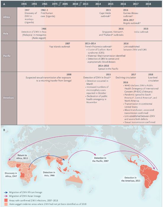
Emergence and Spread of ZIKV and Timeline of the Zika Virus Pandemic.
Panel A shows the major epidemiologic events in the emergence and spread of Zika virus (ZIKV) from its discovery in 1947 through 2018, including outbreaks during which cases of ZIKV-associated birth defects were identified in newborns (*). Panel B is a map of regions where confirmed cases of ZIKV infections have occurred from 2007 through 2018 (red) and areas where Aedes aegypti is endemic but where ZIKV had not yet been identified (pink) as of 2018. Also shown is the migration of African (blue arrow) and Asian (purple arrows) lineages of ZIKV during its global emergence. The epidemiologic events associated with the spread of ZIKV are described in detail in Figure S1 and Table S1 in the Supplementary Appendix.
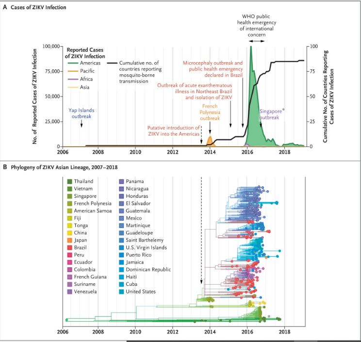
Reported Cases and Spread of the Virus during the Zika Pandemic.
Panel A shows the number of reported cases of ZIKV infection in the Americas, Pacific Islands, Africa, and Asia, as well as the cumulative number of countries or territories worldwide that reported mosquito-borne transmission from January 2007 through December 2018. Panel B shows a ZIKV time-resolved phylogenetic tree that was reconstructed by Nextstrain (https://nextstrain.org/zika. opens in new tab, with permission from Trevor Bedford and Richard Neher) with the use of 506 genomes from 32 countries sampled from February 2013 to September 2017. The American subclade emerged from the Asian lineage and caused outbreaks throughout the Pacific Islands and the epidemic in the Americas.6 The dashed line shows the estimated period (May through November 2013) when ZIKV was introduced into the Americas.8 The cluster of sequences that were obtained from outbreaks in Singapore in August 2016 and Thailand and Vietnam in 2016 and 2017, which are indicated by an asterisk, are distinct from the sequences of pandemic strains from the Pacific Islands and the Americas.
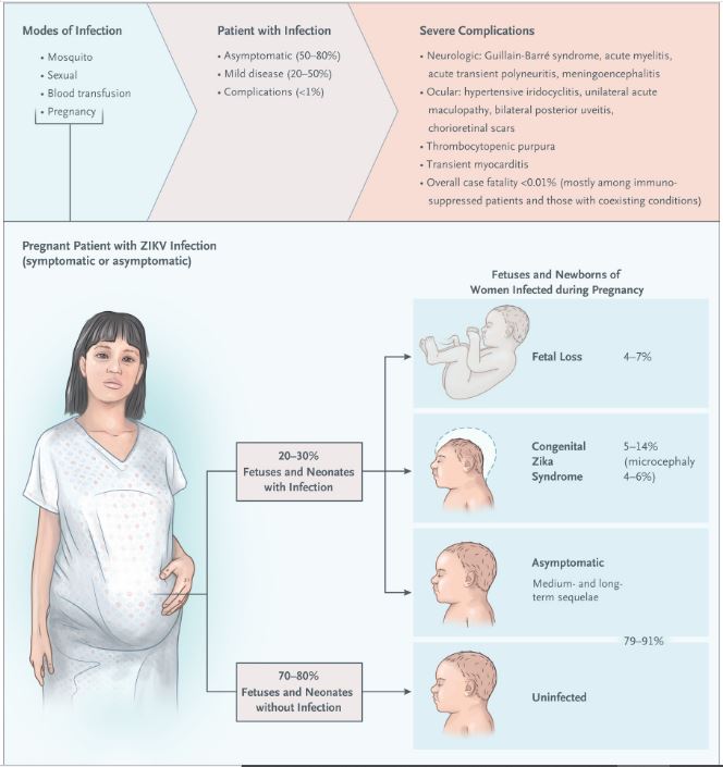
Zika Virus Transmission and Clinical Features.
Shown are the modes of transmission, complications observed in adults and children after infection, and natural history of ZIKV infection during gestation and birth. Percentages of maternal–fetal transmission, fetal loss, acquisition of congenital Zika syndrome, and ZIKV-associated microcephaly among fetuses and infants of women infected with ZIKV during pregnancy were estimated on the basis of the findings of prospective studies and case series (included in the Supplementary Appendix). The estimates do not include data from a prospective study from Rio de Janeiro that showed a high percentage (42%) of adverse outcomes among fetuses and newborns whose mothers were infected with ZIKV.18 At present, the spectrum and risk of medium- and long-term sequelae, including neurodevelopmental delay, have not been fully delineated.
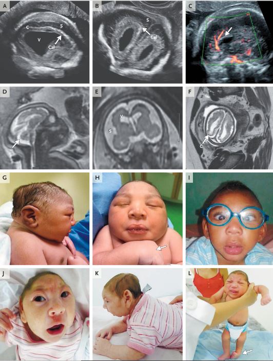
Clinical and Imaging Findings of Congenital Zika Syndrome.
Images illustrate selected features18,46,54-59 of the manifestation of congenital Zika syndrome in fetuses and newborns. Prenatal neurosonograms of fetuses with ZIKV infection (Panels A, B, and C, at 22, 22, and 26 weeks of gestation, respectively) show linear calcifications (Cal, arrows, Panels A and B), increased pericerebral spaces (S, Panels A and B), ventriculomegaly (V, Panel A), cortical thinning (C, Panel A), and dysgenesis of the corpus callosum (arrow, Panel C). MRIs of a fetus with ZIKV infection at 32 weeks of gestation (Panels D, E, and F) show microcephaly (Panel D), hypoplasia of the cerebellum and vermis (arrow, Panel D), premature closure of the fontanels and partial collapse of the skull (Panel D), increased pericerebral spaces (S, Panel E), ventriculomegaly (V, Panel E), cortical thinning (C, Panel F), and dysgenesis of the corpus callosum and gyral anomalies (Panel E). Photographs of three infants — one at 1 week of age (Panels G and H) and 10 months of age (Panel I); the second at 14 days of age (Panels J and K); and the third at 51 days of age (Panel L) — show findings of severe disproportionate microcephaly and cranial dysmorphism (Panels G through L), arthrogryposis (arrow, Panel H), strabismus (Panel I), neck rigidity caused by axial hypertonicity (Panel K), and talipes equinovarus (arrow, Panel L).
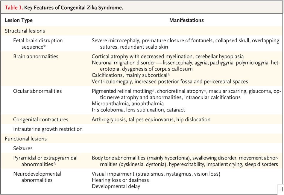
Key Features of Congenital Zika Syndrome
The condition of the woman, say doctors, is stable. She delivered on June 7. The patient was diagnosed and hospitalised at KIMS. Samples of 13 persons, suspected to be positive for Zika virus, have been sent to the National Institute of Virology in Pune. The symptoms of Zika virus include fever, skin rashes, conjunctivitis, muscle and joint pain, malaise, and headache.
According to WHO : Zika virus is primarily transmitted by the bite of an infected mosquito from the Aedes genus, mainly Aedes aegypti, in tropical and subtropical regions. Aedes mosquitoes usually bite during the day, peaking during early morning and late afternoon/evening. This is the same mosquito that transmits dengue, chikungunya and yellow fever.
Zika virus is also transmitted from mother to fetus during pregnancy, through sexual contact, transfusion of blood and blood products, and organ transplantation.
In October 2015, Brazil reported an association between Zika virus infection and microcephaly. Outbreaks and evidence of transmission soon appeared throughout the Americas, Africa, and other regions of the world. To date, a total of 86 countries and territories have reported evidence of mosquito-transmitted Zika infection.
No vaccine is yet available for the prevention or treatment of Zika virus infection. Development of a Zika vaccine remains an active area of research. The virus was first isolated in 1947 in Uganda's zika forest.
The virus is spread through the Aedes species of mosquitoes, which is found in high density across the state. Aedes mosquitoes, which are also carriers of dengue, breed in stagnant freshwater and rest mostly indoors.
Zika virus has often been linked to birth defects and development of Guillain-Barre syndrome, where one's immune system attacks the nerves. Some people infected by it might not show any signs or symptoms. However, in pregnant women the infection can seriously harm the developing foetus and lead to congenital anomalies.
Zika Virus Pandemic
Emergence and Spread of ZIKV and Timeline of the Zika Virus Pandemic.
Panel A shows the major epidemiologic events in the emergence and spread of Zika virus (ZIKV) from its discovery in 1947 through 2018, including outbreaks during which cases of ZIKV-associated birth defects were identified in newborns (*). Panel B is a map of regions where confirmed cases of ZIKV infections have occurred from 2007 through 2018 (red) and areas where Aedes aegypti is endemic but where ZIKV had not yet been identified (pink) as of 2018. Also shown is the migration of African (blue arrow) and Asian (purple arrows) lineages of ZIKV during its global emergence. The epidemiologic events associated with the spread of ZIKV are described in detail in Figure S1 and Table S1 in the Supplementary Appendix.
Reported Cases and Spread of the Zika Virus
Reported Cases and Spread of the Virus during the Zika Pandemic.
Panel A shows the number of reported cases of ZIKV infection in the Americas, Pacific Islands, Africa, and Asia, as well as the cumulative number of countries or territories worldwide that reported mosquito-borne transmission from January 2007 through December 2018. Panel B shows a ZIKV time-resolved phylogenetic tree that was reconstructed by Nextstrain (https://nextstrain.org/zika. opens in new tab, with permission from Trevor Bedford and Richard Neher) with the use of 506 genomes from 32 countries sampled from February 2013 to September 2017. The American subclade emerged from the Asian lineage and caused outbreaks throughout the Pacific Islands and the epidemic in the Americas.6 The dashed line shows the estimated period (May through November 2013) when ZIKV was introduced into the Americas.8 The cluster of sequences that were obtained from outbreaks in Singapore in August 2016 and Thailand and Vietnam in 2016 and 2017, which are indicated by an asterisk, are distinct from the sequences of pandemic strains from the Pacific Islands and the Americas.
Zika Virus Transmission and Clinical Features
Zika Virus Transmission and Clinical Features.
Shown are the modes of transmission, complications observed in adults and children after infection, and natural history of ZIKV infection during gestation and birth. Percentages of maternal–fetal transmission, fetal loss, acquisition of congenital Zika syndrome, and ZIKV-associated microcephaly among fetuses and infants of women infected with ZIKV during pregnancy were estimated on the basis of the findings of prospective studies and case series (included in the Supplementary Appendix). The estimates do not include data from a prospective study from Rio de Janeiro that showed a high percentage (42%) of adverse outcomes among fetuses and newborns whose mothers were infected with ZIKV.18 At present, the spectrum and risk of medium- and long-term sequelae, including neurodevelopmental delay, have not been fully delineated.
Congenital Zika Virus Syndrome
Clinical and Imaging Findings of Congenital Zika Syndrome.
Images illustrate selected features18,46,54-59 of the manifestation of congenital Zika syndrome in fetuses and newborns. Prenatal neurosonograms of fetuses with ZIKV infection (Panels A, B, and C, at 22, 22, and 26 weeks of gestation, respectively) show linear calcifications (Cal, arrows, Panels A and B), increased pericerebral spaces (S, Panels A and B), ventriculomegaly (V, Panel A), cortical thinning (C, Panel A), and dysgenesis of the corpus callosum (arrow, Panel C). MRIs of a fetus with ZIKV infection at 32 weeks of gestation (Panels D, E, and F) show microcephaly (Panel D), hypoplasia of the cerebellum and vermis (arrow, Panel D), premature closure of the fontanels and partial collapse of the skull (Panel D), increased pericerebral spaces (S, Panel E), ventriculomegaly (V, Panel E), cortical thinning (C, Panel F), and dysgenesis of the corpus callosum and gyral anomalies (Panel E). Photographs of three infants — one at 1 week of age (Panels G and H) and 10 months of age (Panel I); the second at 14 days of age (Panels J and K); and the third at 51 days of age (Panel L) — show findings of severe disproportionate microcephaly and cranial dysmorphism (Panels G through L), arthrogryposis (arrow, Panel H), strabismus (Panel I), neck rigidity caused by axial hypertonicity (Panel K), and talipes equinovarus (arrow, Panel L).
Key Features of Congenital Zika Syndrome
Key Features of Congenital Zika Syndrome
Kerala, which has been reporting a surge in coronavirus cases, has officially confirmed its first case of Zika virus - a mosquito-borne viral infection - after the virus was detected in the blood samples of a 24-year-old pregnant woman in Thiruvananthapuram. pic.twitter.com/gn7qRhF5dC
— VISUM Expresso (@VisumExpresso) July 9, 2021

ആരാണ് കേരളത്തിലെ പട്ടിണി മാറ്റിയത്, 2024 ലോക്സഭാ തിരഞ്ഞെടുപ്പ് അടുക്കുമ്പോൾ ചിന്തിക്കേണ്ട ചില കാര്യങ്ങൾ
Updated:
March 28, 2024 , 01:17 PM


കരിപ്പൂരിൽ വൻ സ്വർണവേട്ട; വിമാന ജീവനക്കാരൻ കരിപ്പൂർ കരുവാംകല്ല് സ്വദേശി മുഹമ്മദ് ഷമീം പിടിയിൽ
Updated:
July 31, 2022 , 08:11 PM


COMMENTS
Be the first to comment
Latest Post

Literature
കടലാഴങ്ങളെ പുണർന്ന നാവികൻ, കടൽ ഒറ്റയ്ക്കു ക്ഷണിച്ചപ്പോൾ- ഒരു വായനാനുഭവം
Saturday, 03 May, 2025


LEAVE A REPLY